Would you like to switch to the accessible version of this site?

Go to accessible site Close modal window

Don't need the accessible version of this site?

Hide the accessibility button Close modal window
Accessibility View Close toolbar
VH

Our Location

(707) 206-0290

Open mobile navigation
  • Home
  • New Patient Center
    • Online Forms
    • Insurance and Payments
  • About Us
    • Meet the Optometrist
  • Eyecare Services
    • Contact Lens Exams
    • Eye and Vision Exams
    • Lasik Eye Surgery
    • Hard to Fit Contacts
    • Computer Vision Treatment
    • Sports Vision
    • Sunglasses
  • Frames
    • Designer Frames
  • Testimonials
  • Contact Us
    • Appointment Request
  • Community Content
  • Home >
  • Articles >
  • Newsletters >
  • How is Eye Pressure Measured?

How is Eye Pressure Measured?

  • Created in Newsletters

Eye doctor doing an exam

Testing Your Eye Pressure

High pressure inside your eyes increases your risk of glaucoma, a disease that can cause vision loss. Although you can't tell when your eye pressure is rising, a simple test conducted at your optometrist's office helps your eye doctor detect and treat pressure changes.

Why It's Important to Keep Your Pressure Under Control

Your eye is filled with a clear fluid called aqueous humor that delivers nutrients to the cornea and lens and removes waste products. The pressure produced by the fluid helps your eyes keep their round shape.

Drainage channels constantly remove old aqueous humor and make room for a new supply of fluid. If the drainage channels become blocked, your eye pressure rises, which can cause optic nerve damage.

The optic nerve carries electrical impulses from your eyes to your brain. Once the impulses reach the brain, they're processed and transformed into images. If the optic nerve becomes damaged by high pressure inside your eyes, the impulses may not reach your brain. This can cause partial or complete vision loss.

Unfortunately, you probably won't notice any symptoms if you have a common type of glaucoma called open-angle glaucoma. Despite the lack of symptoms, open-angle glaucoma can slowly damage your vision. Angle-closure glaucoma, the more severe form of the condition, occurs suddenly and does cause symptoms. Symptoms of angle-closure glaucoma include pain, sudden loss of vision, nausea, headache, halos around lights, and blurry vision.

So how high is too high when it comes to eye pressure? The American Academy of Ophthalmology reports that pressure between 10 and 21 millimeters of mercury (mmHg) is normal and anything above that is too high. Some people who have eye pressure that's technically in the normal range may also be at risk of developing glaucoma.

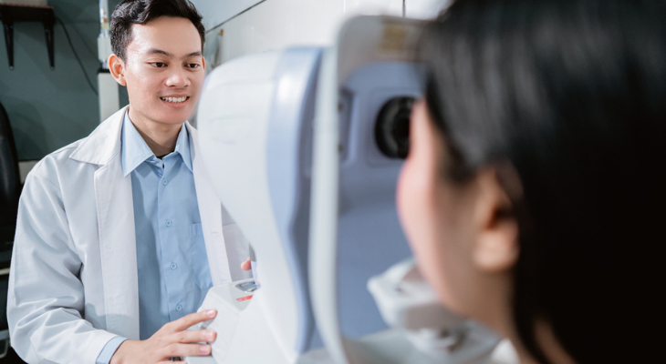
3 Ways to Test Eye Pressure

Eye doctors use several methods to test eye pressure, including:

  • Non-Contact Tonometry. Commonly called the "air puff" test, non-contact tonometry measures how much your corneas flatten when exposed to a burst of air. While you look at a light, a puff of air flattens your cornea. Generally, the more force needed to flatten the cornea, the higher the pressure inside your eye.
  • Goldmann Applanation Tonometry. Before this test begins, your eye doctor puts drops in your eyes that numb them and also adds a few drops of a blue dye. After your eye is numb, your doctor looks in your eyes with a special machine called a slit-lamp microscope. You'll notice a glowing blue light that makes the dye added to your eyes visible. The test is conducted by pressing the tip of a small probe against your cornea. The tonometer measures how much pressure it takes to flatten the cornea.
  • Electronic Tonometry. An electronic tonometer looks a little like a pen. During the test, the device is briefly held against your eye after it's numbed with eye drops. Your eye pressure reading appears on a small screen on the device.

What Happens If Your Pressure Is High

A higher-than-normal pressure reading doesn't necessarily mean that you have glaucoma. Some people naturally have higher pressure readings than others, yet have no damage to their optic nerves.

If you have a high eye pressure reading, your eye doctor will take a close look at your optic nerve. Dilating your pupil with special drops makes it possible to view the optic nerve and spot damage. They may also take a few pictures of the nerve. In addition to checking out your optic nerve, your eye doctor measures the thickness of your corneas. A thick cornea could make your pressure measurement seem higher than it really is. As part of your examination, your eye doctor will also test your side vision and check to see if the drainage channels inside your eye are blocked.

If your pressure is too high, you may need one or more of these glaucoma treatments:

  • Eye drops to decrease pressure
  • Laser eye surgery to improve drainage
  • Traditional eye surgery if laser therapy isn't helpful

Annual eye examinations and eye pressure tests help you protect your eyesight and ensure that you get the treatment you need should you ever develop glaucoma. Contact us to schedule your next comprehensive eye examination.

Sources:

Bright Focus Foundation: How Is Eye Pressure Measured?

American Academy of Ophthalmology: Eye Pressure Testing, 2/26/18

All About Vision: Do I Have to Get the Air Puff Test, 10/21

Glaucoma Research Foundation: The Importance of Corneal Thickness, 10/29/17

  • Common Eye Conditions
    • Age-Related
    • Cognitive and Acquired
    • Vision Impairment
    • Injury & Irritation
    • Eye Lids
    • Changes in Appearance
  • Contact Lenses
  • Eye Diseases
  • Eyeglasses
    • Eyeglass Lenses
  • Eye Symptoms
  • How the Eyes Work
    • Basic Visual Skills
  • Pediatric Vision
  • Protecting Your Eyes
  • Visual Rehabilitation
  • Vision Problems
  • What is Vision Therapy
  • Vision Therapy Programs
  • We Can Help With
    • Cataracts
    • Corneal Disorders
      • Disorders
    • Glaucoma
    • Refractive Disorders
    • Adult Strabismus
    • Retinal Disorders
  • Newsletters
    • Glasses & Frames
    • Contacts
    • Tips for Healthy Eyes
    • Conditions That Affect Vision
    • Kid's Vision
    • Amazing, Interesting Eyes
    • Medical Perspectives

Contact Us

We look forward to hearing from you.

Hours of Operation

Our Regular Schedule

Rohnert Park Office

Monday:

11:00 am-4:00 pm

Tuesday:

10:00 am-7:00 pm

Wednesday:

9:00 am-7:00 pm

Thursday:

9:00 am-5:00 pm

Friday:

9:00 am-5:00 pm

Saturday:

Closed

Sunday:

Closed

Locations

Find us on the map

    No testimonials found. Please add

Featured Articles

  • March Newsletter: Are Your Eyes Getting Enough Vitamin D?

    Not getting enough of a key vitamin might increase your risk of several eye diseases. ...

    Read More
  • March Newsletter: Is Your Prescription Up to Date? What You Need to Know about When and Why You should See Your Optometrist

    Not sure when you need to see the eye doctor? ...

    Read More
  • Saccadic Fixators

    This entails a wall-mounted square board with a starburst design. Along the various striations of the starburst are lighted buttons. As the buttons light up, the patient works quickly to see how many of these lit buttons they can push before they go out. The key is to keep the head still. Peripheral ...

    Read More
  • Patches

    Eye patches are used to strengthen muscle control in weak eyes. By placing a patch over the strong eye, the weaker eye is forced to do the heavy lifting. While it may be uncomfortable for the patient at first, the muscle controlling the weaker eye will become tougher and more resilient. This will allow ...

    Read More
  • Rotation Trainers

    Rotation trainers consist of a disk – with various designs – that is attached to a rod-like base. As the disk rotates, the patient is asked to perform tasks that are designed to test and enhance eye-hand coordination, space awareness, perceptual awareness and visual acuity. ...

    Read More
  • Computer Software

    Computer aided vision therapy consists of a software package designed to enhance eye tracking skills, visual thinking, processing skills and binocular vision skills. Eye teaming, focusing and tracking are not optical in nature, and problems in these areas are the result of poor eye muscles. Specialized ...

    Read More
  • Prisms

    A prism has the same cross-section across the entire length of its shape. When used in eyeglasses, they often correct abnormalities associated with nearsightedness, farsightedness and double vision. Eyeglasses, or corrective lenses, reduce or increase the size of the image based on the eyes’ ability ...

    Read More
  • February Newsletter: Age-Related Macular Degeneration (AMD) Awareness Month

    Do you know how age-related macular degeneration could affect your vision? ...

    Read More
  • February Newsletter: Why Dry Eye Is So Difficult to Treat

    Are dry eyes a frequent problem? Try these tips. ...

    Read More
  • January Newsletter: How to Tell if You Have Macular Degeneration

    Do you know the symptoms that may mean you have macular degeneration? ...

    Read More

Newsletter Signup

Sign up for more articles!

  • Copyright © 2023 MH Sub I, LLC dba iMatrix
  • Admin Log In
  • Site Map