Would you like to switch to the accessible version of this site?

Go to accessible site Close modal window

Don't need the accessible version of this site?

Hide the accessibility button Close modal window
Accessibility View Close toolbar
VH

Our Location

(707) 206-0290

Open mobile navigation
  • Home
  • New Patient Center
    • Online Forms
    • Insurance and Payments
  • About Us
    • Meet the Optometrist
  • Eyecare Services
    • Contact Lens Exams
    • Eye and Vision Exams
    • Lasik Eye Surgery
    • Hard to Fit Contacts
    • Computer Vision Treatment
    • Sports Vision
    • Sunglasses
  • Frames
    • Designer Frames
  • Testimonials
  • Contact Us
    • Appointment Request
  • Community Content
  • Home >
  • Articles >
  • How the Eyes Work >
  • Eye Anatomy 101

Eye Anatomy 101

  • Created in How the Eyes Work

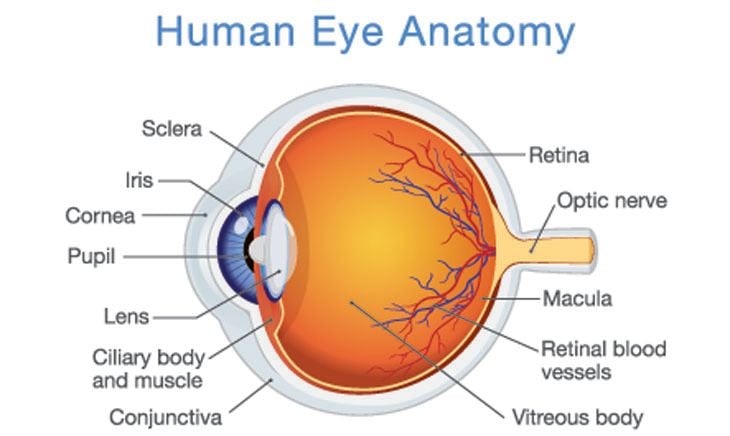
Human Eye Anatomy

The eye has many parts that work together to create vision. The eyes themselves are only part of “seeing.” The brain is also involved. The eyes, though, begin the complex process of vision by gathering, focusing and passing on visual information to the brain.

To help you learn how the eye works, here is a quick overview of the anatomy of the eye. This information is also useful in understanding which parts of the eye are affected by different health problems of the eye.

Cornea. Light first enters the eye through this transparent, dome-shaped surface that covers the front of the eye. The cornea bends — or refracts — the light onto the eye’s lens.

Pupil. This is the round hole at the front of the eye that appears black. It is located behind the middle of the cornea and is surrounded by the iris.

Iris. The iris, or the colored part of the eye surrounding the pupil, controls how much light enters the eye. The iris can make the pupil bigger or smaller by opening or closing.

Lens. Behind the pupil and the iris is a transparent structure that looks similar in shape to the lens of a magnifying glass. Unlike glass lenses, though, this part of the eye can change shape. This enables it to bend the rays of light even more, so they land in the right place on the retina, at the back of the eye.

Vitreous body, or gel. The eye is filled with a gel that helps it keep its round shape. Light entering the eye first passes through the cornea then the lens and then the vitreous body before reaching the retina.

Retina. At the back of the eye is the retina, or a thin layer of light-sensitive nerve cells. The retina contains different types of photoreceptors — called rods and cones — which respond to light that lands on them.

Cones. These light-sensitive nerve cells are located in the macula, which is located at the center of the retina. Cones are necessary for focused central vision. Cones also enable you to see colors in bright-light conditions.

Rods. These light-sensitive nerve cells surround the macula and extend to the edge of the retina. The rods provide you with your side, or peripheral, vision. They also help you see at night and in dim light.

Optic nerve. The cells of the retina turn light into electrical impulses. These electrical signals are collected by the optic nerve — a bundle of about 1 million nerve fibers — and transmitted to the brain. The brain puts all this information together to produce the image that you see.

To learn more about eye anatomy or to have your eye health examined, please contact our practice.

  • Common Eye Conditions
    • Age-Related
    • Cognitive and Acquired
    • Vision Impairment
    • Injury & Irritation
    • Eye Lids
    • Changes in Appearance
  • Contact Lenses
  • Eye Diseases
  • Eyeglasses
    • Eyeglass Lenses
  • Eye Symptoms
  • How the Eyes Work
    • Basic Visual Skills
  • Pediatric Vision
  • Protecting Your Eyes
  • Visual Rehabilitation
  • Vision Problems
  • What is Vision Therapy
  • Vision Therapy Programs
  • We Can Help With
    • Cataracts
    • Corneal Disorders
      • Disorders
    • Glaucoma
    • Refractive Disorders
    • Adult Strabismus
    • Retinal Disorders
  • Newsletters
    • Glasses & Frames
    • Contacts
    • Tips for Healthy Eyes
    • Conditions That Affect Vision
    • Kid's Vision
    • Amazing, Interesting Eyes
    • Medical Perspectives

Contact Us

We look forward to hearing from you.

Hours of Operation

Our Regular Schedule

Rohnert Park Office

Monday:

11:00 am-4:00 pm

Tuesday:

10:00 am-7:00 pm

Wednesday:

9:00 am-7:00 pm

Thursday:

9:00 am-5:00 pm

Friday:

9:00 am-5:00 pm

Saturday:

Closed

Sunday:

Closed

Locations

Find us on the map

    No testimonials found. Please add

Featured Articles

  • March Newsletter: Are Your Eyes Getting Enough Vitamin D?

    Not getting enough of a key vitamin might increase your risk of several eye diseases. ...

    Read More
  • March Newsletter: Is Your Prescription Up to Date? What You Need to Know about When and Why You should See Your Optometrist

    Not sure when you need to see the eye doctor? ...

    Read More
  • Saccadic Fixators

    This entails a wall-mounted square board with a starburst design. Along the various striations of the starburst are lighted buttons. As the buttons light up, the patient works quickly to see how many of these lit buttons they can push before they go out. The key is to keep the head still. Peripheral ...

    Read More
  • Patches

    Eye patches are used to strengthen muscle control in weak eyes. By placing a patch over the strong eye, the weaker eye is forced to do the heavy lifting. While it may be uncomfortable for the patient at first, the muscle controlling the weaker eye will become tougher and more resilient. This will allow ...

    Read More
  • Rotation Trainers

    Rotation trainers consist of a disk – with various designs – that is attached to a rod-like base. As the disk rotates, the patient is asked to perform tasks that are designed to test and enhance eye-hand coordination, space awareness, perceptual awareness and visual acuity. ...

    Read More
  • Computer Software

    Computer aided vision therapy consists of a software package designed to enhance eye tracking skills, visual thinking, processing skills and binocular vision skills. Eye teaming, focusing and tracking are not optical in nature, and problems in these areas are the result of poor eye muscles. Specialized ...

    Read More
  • Prisms

    A prism has the same cross-section across the entire length of its shape. When used in eyeglasses, they often correct abnormalities associated with nearsightedness, farsightedness and double vision. Eyeglasses, or corrective lenses, reduce or increase the size of the image based on the eyes’ ability ...

    Read More
  • February Newsletter: Age-Related Macular Degeneration (AMD) Awareness Month

    Do you know how age-related macular degeneration could affect your vision? ...

    Read More
  • February Newsletter: Why Dry Eye Is So Difficult to Treat

    Are dry eyes a frequent problem? Try these tips. ...

    Read More
  • January Newsletter: How to Tell if You Have Macular Degeneration

    Do you know the symptoms that may mean you have macular degeneration? ...

    Read More

Newsletter Signup

Sign up for more articles!

  • Copyright © 2023 MH Sub I, LLC dba iMatrix
  • Admin Log In
  • Site Map